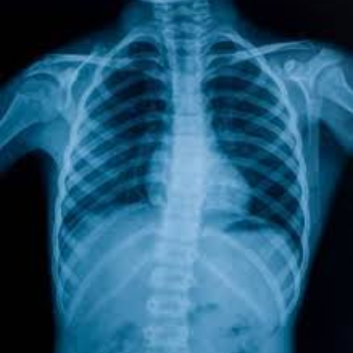
A solid understanding of chest anatomy is the cornerstone of professional radiology practice. This video is designed to build a clear, practical foundation for radiology technicians and specialists who want to move beyond memorization and toward true clinical understanding.
What You Will Learn in This Video
In this session, you will gain a structured and practical understanding of:
-
The essential chest anatomy every radiology technician and specialist must know
-
A simple and logical explanation of the thoracic circulatory system
-
How blood moves within the chest and why this movement is critical in radiological imaging
-
The basics of reading and describing chest X-rays with confidence
-
How to connect anatomy, imaging appearance, and patient positioning
-
Developing the correct radiological way of thinking, not just memorizing information
This approach helps you see anatomy as it appears on images, not just as textbook diagrams.
Who Is This Video For?
This video is ideal for:
-
Students and graduates of health institutes and medical colleges
-
Beginner radiology technicians or specialists at the start of their training
-
Anyone who wants to master the correct fundamentals before progressing to contrast studies and advanced imaging
If you feel confused when looking at chest images or struggle to connect anatomy with radiological findings, this video is made for you.
The Main Goal of the Video
The primary goal is to help you understand the circulatory system as a complete functional unit, forming a strong base that can be expanded later.
This foundation is essential for:
-
Advanced contrast examinations
-
CT scan Level 2 studies
-
Confident and safe use of contrast agents
By the end of this video, you will be able to visualize any patient entering the imaging room, understand what you are seeing, and apply contrast studies with purpose—not guesswork.
From Memorization to True Understanding
Even if you struggle with memorizing pathology or are still at a beginner level, this video will help you:
-
Describe abnormalities accurately
-
Identify their location within the chest
-
Think logically about what you see on the image and why it appears that way
This is the first step toward becoming a radiology professional who understands images, not just looks at them.