Lower limb radiography is a core skill in daily radiology practice. This video is designed to remove confusion and provide a clear, structured understanding of lower limb bone X-rays, focusing on anatomy, positioning, and correct image interpretation.
What You Will Learn in This Video
In this video, you will develop a solid and practical understanding of:
-
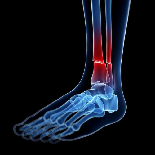
Essential lower limb bone anatomy as seen in radiographic imaging
-
The relationship between bones within each joint and how this affects image interpretation
-
The impact of patient positioning and X-ray beam angle on the final image shape
-
A systematic approach to describing lower limb radiographs
-
How to identify and describe obvious abnormalities and changes confidently
This step-by-step method helps you read images logically instead of guessing.
Who Is This Video For?
This video is suitable for:
-
Students and graduates of health institutes and medical colleges
-
Radiology technicians and specialists in daily clinical practice
-
Anyone who feels that lower limb X-rays look similar or confusing
If you struggle to differentiate structures or feel unsure while describing images, this video will give you a clear framework.
The Goal of the Video
The main goal is to help you master the basics of lower limb radiography.
This foundation allows you to later:
-
Handle more practical and clinical cases
-
Link plain X-ray findings to CT scans
-
Progress confidently to advanced imaging levels, including:
-
MRI
-
Evaluation of ligament injuries
-
Assessment of osteoarthritis
-
Build Strong Foundations for Advanced Imaging
By the end of this video, you will understand what you are looking at, why it appears that way, and how to describe it accurately—setting you up for success in advanced radiology studies.