Upper limb bone radiography is a fundamental skill for radiology technicians and specialists. This video focuses on building a clear and practical understanding of upper limb X-rays, helping you move from confusion to confident image interpretation.
What You Will Learn in This Video
This video provides a structured introduction to upper limb bone radiography, including:
-
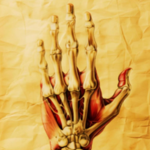
Essential anatomy of the upper limb bones as required in daily radiology practice
-
The relationship between bones, joints, and imaging angles
-
How incorrect positioning can significantly change image appearance
-
An introduction to the descriptive language used in bone radiography
-
How to differentiate normal findings from changes that require attention
This approach helps you describe images logically rather than relying on memorization.
Who Is This Video For?
This video is ideal for:
-
Students and graduates of health institutes and medical colleges
-
Radiology technicians and specialists at the beginning of their careers or training
-
Anyone who finds bone radiographs confusing or too similar
If you struggle to recognize structures or describe findings confidently, this video is designed for you.
The Goal of the Video
The primary goal is to build a strong foundation in bone radiography.
This foundation allows you to:
-
Progress to more complex cases and advanced applications
-
Understand how plain X-rays connect to CT and MRI imaging
-
Learn how to plan imaging techniques and interpret studies using radiology software
Preparing for Advanced Imaging Levels
By mastering the basics presented in this video, you will be ready to move forward with confidence into advanced imaging techniques, supported by a clear understanding of anatomy, positioning, and image interpretation.